
Identifying problematic veins is an essential clinical skill for every nurse involved in IV therapy. Proper vein assessment not only reduces patient discomfort but also increases the success rate of cannulation on the first attempt. With practice, clinical reasoning and the support of cannulation courses for nurses, you can become more confident in recognising challenging veins and selecting the best approach.
This guide explains how to identify problematic veins, what signs to look for, and professional techniques to improve cannulation success in a UK healthcare setting.
What Makes a Vein Difficult to Cannulate?
Several factors can influence vein accessibility. Recognising these early helps you plan your technique effectively.
Common Characteristics of Difficult Veins
- Veins that roll, collapse or move under the skin
- Thin, fragile or hardened veins
- Deep veins are located beneath adipose tissue
- Dehydrated patients with reduced vascular volume
- Oedematous limbs
- History of chemotherapy, resulting in vein scarring
- Patients with frequent cannulations, causing vein exhaustion
Patient Groups Likely to Have Difficult Veins
Some patient populations naturally present a challenge. Continually assess carefully when working with:
- Elderly patients – fragile, thin and tortuous veins
- Paediatric patients – tiny and fast-collapsing veins
- Oncology patients – long-term vein damage
- IV drug users – scarred or inaccessible veins
- Obese patients – deeper veins with poor visibility
- Dehydrated or shocked patients – minimal venous filling
Attending high-quality cannulation courses for nurses enhances your ability to safely and effectively manage these higher-risk patient groups.
How to Identify a Good vs Difficult Vein
✔️ Signs of a Good Vein
- Soft and bouncy on palpation
- Well-filled and visible after tourniquet application
- Straight pathway with minimal branching
- No hardness, scarring or valve clusters
❌ Signs of a Difficult Vein
- Feels hard, fibrotic or rope-like
- Does not refill when pressure is released
- Rolls away during palpation
- Appears flat, narrow or poorly defined
- Has visible valve bulges
- Painful when touched
Techniques to Improve Vein Identification
1. Use Both Vision and Palpation
Do not rely solely on your eyes. Some excellent veins are not visible but feel bouncy under the skin. Palpation helps detect depth, size and direction.
2. Lower the Limb
Gravity increases venous filling. Ask the patient to lower their arm below heart level for 30–60 seconds.
3. Warm the Area
Applying a warm compress for 5–10 minutes encourages vasodilation and vein expansion.
4. Hydrate the Patient (if appropriate)
If the patient is not fluid-restricted, encourage oral fluids or request IV hydration before attempting cannulation.
5. Use a Proper Tourniquet Technique
A correctly tightened tourniquet should:
- Engorge the vein
- Allow arterial flow
- Make veins easier to palpate
Avoid overtightening, as it can collapse fragile veins.
6. Tap and Massage
Gentle tapping or stroking along the vein encourages dilation and filling. Avoid aggressive tapping as it may cause bruising. if you want more career boosting blog here.
7. Ask the Patient to Make a Fist
This increases venous pressure and makes superficial veins more prominent.
Advanced Tools for Identifying Difficult Veins
Vein Locators
Infrared vein finders can help visualise deeper or hard-to-locate veins, especially in paediatrics or obese patients.
Ultrasound Guidance
Ultrasound-guided cannulation is increasingly used in UK hospitals for:
- Deep veins
- Poor venous access
- Emergencies
Many hospitals now offer training sessions alongside structured cannulation education.
Common Veins to Use – and Avoid
Veins to Favour
- Dorsal hand veins – accessible but can be painful
- Cephalic vein – straight, consistent pathway
- Basilic vein – a larger vein, often underused
- Median cubital vein – excellent for confident cannulators
Veins to Avoid
- Areas around joints (movement increases failure)
- Veins that feel cord-like or hardened
- Veins close to infection, wounds, bruising
- Previously used or recently cannulated sites

Professional Tips for Improving Cannulation Success
Before the Attempt
- Explain the procedure to reduce anxiety and muscle tension
- Position the patient comfortably
- Prepare all equipment in advance
During the Attempt
- Stretch the skin to stabilise the vein
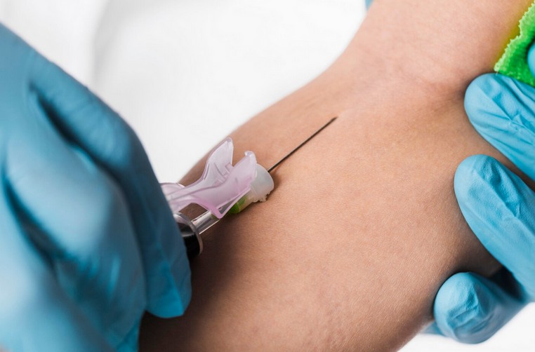
- Insert the cannula bevel-up at the correct angle (usually 10–30°)
- Watch for flashbacks and advance gently
If the Vein Rolls
- Anchor firmly below the insertion point
- Use your non-dominant hand for stabilisation
If the Vein Collapses
- Reduce tourniquet tension
- Choose a larger vein or a different site
When to Stop and Reassess
Stop immediately if:
- The patient reports sharp pain
- Bruising forms
- Swelling appears
- The vein disappears despite repeated attempts
Never attempt cannulation more than twice before asking a senior or specialist for support.
Conclusion
Identifying problematic veins is a skill refined through practice, proper assessment and continuous training. By understanding patient factors, using effective vein-finding techniques and applying evidence-based clinical methods, nurses can significantly improve their cannulation success rate. Professional development, including cannulation courses for nurses, empowers healthcare staff to handle complex venous access scenarios with confidence and competence.
FAQs
1. What are the signs of a problematic vein?
Hardness, rolling, poor refill, fragility and low visibility are common indicators.
2. How can I make a vein easier to see?
Warmth, gravity, hydration and correct tourniquet use help veins fill.
3. Should I attempt cannulation more than twice?
No. Follow UK guidelines—after two failed attempts, seek assistance.
4. Why do elderly patients have problematic veins?
Their veins are fragile, thin and easily collapse under pressure.
5. Are vein finders helpful?
Yes. Infrared devices and ultrasound are excellent for locating deep or hidden veins